

In a study directed and coordinated by the Department of Information Engineering and the Department of Medical-Surgical Specialties, Radiological Sciences and Public Health of the University of Brescia, a group of researchers studied, for the first time internationally, the prognostic capacity of the Artificial Intelligence techniques (Deep Learning and Computer Vision) starting from the analysis of the radiographic images of the chest (CXR) used in the estimate of the degree of severity of Covid-19 pneumonia.
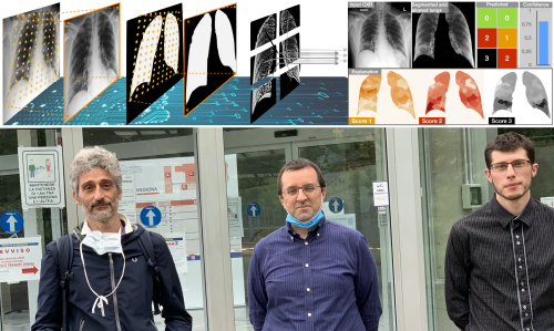
The companies Philips Italia and El.Co and the IT team of the ASST Spedali Civili of Brescia were involved in the recovery of the data. From the analysis of several thousand images, corresponding to the complete flow of radiographs of Covid-19 patients admitted from the beginning of March to the beginning of April at the Asst Spedali Civili of Brescia – an order of magnitude greater than the number of publicly available databases – BrixIA-Net , The new Deep Learning architecture designed by the University of Brescia, was able to describe the degree of pulmonary impairment of the patients (interstitial and alveolar), based on a CXR score system expressed in six values (corresponding to as many lung areas), indicating a progressive severity of pneumonia.
This evaluation system, called Brixia-score, was developed by the doctors of the Radiology 2 Operative Unit directed by Prof. Maroldi and has been integrated into daily clinical activity since the days when the pressure on the hospital wards, and consequently on the Radiologies of the ASST Spedali Civili, was greater. The BrixIA-Net network, suitably trained on a vast set of radiographic images and Brixia-score reporting data, as well as on other public radiographic databases, has proven to be an extremely effective aid in the diagnosis and assessment of the severity of Covid’s pneumonia. 19.
«In light of the recorded performances, equal to or even superior to those of individual radiologists – explains prof. Alberto Signoroni of the Information Engineering Department, scientific manager of the study – our study supports the possibility of using this tool in collaborative human-machine scenarios for computer-aided monitoring of disease progression. In fact, with a new technique designed specifically for this case study, highly resolute maps of “explainability” are generated, that is visual feedback that allows the radiologist to better evaluate both the suggestions and any errors of the machine ».
“In order to carry out all the phases, from the identification and mapping of the lungs to the evaluation of the degree of compromise in the various areas, we have designed an architecture that combines several state-of-the-art technologies in a single model” – observes the new doctor research Mattia Savardi, who oversaw the development of the software. “The application of deep learning and computer vision techniques in the area of diagnostic imaging is nothing new. The element of novelty – declares prof. Davide Farina of the Department of Medical-Surgical Specialties, Radiological Sciences and Public Health – is represented by the fact that artificial intelligence, in this case, provides the radiologist doctor not so much a diagnosis as a support in defining the severity of the disease, which is also evaluated on a first level exam. This means being able to minimize the subjective element of the evaluation and speed up reporting times, making workflow more efficient “.